Nessun risultato di contenuto corrisponde alla tua parola chiave.
Contenuto

Sei uscito con successo.


Non sei ancora registrato?
Aiutare a prevenire e trattare le ulcere del piede diabetico
La sindrome del piede diabetico (DFS) è una complicanza comune del diabete mellito che colpisce un quarto dei pazienti affetti da diabete in un certo momento della loro vita. Il rischio di sviluppare DFS aumenta con il passare del tempo.
Una delle conseguenze è la lesione cutanea o l'ulcerazione del piede, nota come ulcera del piede diabetico (DFU). Questa condizione comprende tutte le alterazioni del piede derivanti dalla polineuropatia diabetica, nonché le alterazioni micro e macroangiopatiche diabetiche.

Cliccando "Confirm" dichiari di essere un operatore sanitario.
Se sei un paziente o un giornalista visita le pagine a te dedicate.
Conferma Sì, sono un operatore sanitario. Cancella No, non sono un operatore sanitario.Infezione nelle ulcere del piede diabetico (DFU)
19- 0%
delle persone affette da diabete svilupperà una DFU nel corso della propria vita.[2]
Più di
0%
di queste si infettano.[3]
Fino a
0%
delle DFU gravemente infette portano all'osteomielite.[4]
Infezione delle ulcere del piede diabetico: Prevenzione & Trattamento
L'infezione nella DFU non solo è associata a una guarigione difficile o ritardata, ma rappresenta anche un fattore di rischio per il ricovero ospedaliero. Inoltre, dal 10% al 45% dei pazienti ricoverati in ospedale per infezioni ai piedi necessita di un nuovo ricovero entro un anno, con conseguente aumento dei costi sanitari. Nei soggetti con infezioni gravi o osteomielite, la probabilità di amputazione aumenta significativamente.[5]
La diagnosi della DFU si basa principalmente sui segni clinici:[5]
E tecniche di esame clinico:[6]
La gravità delle infezioni del piede diabetico è classificata secondo la classificazione IWGDF/IDSA:[6]

Nessun sintomo o segno sistemico o locale di infezione.
Pulire con soluzioni detergenti per ferite e sbrigliare.

Infetto: Sono presenti almeno due di queste condizioni: gonfiore o indurimento locale; eritema >0,5 ma <2 cm intorno alla ferita; dolorabilità o dolore locale; aumento locale della temperatura; secrezione purulenta.
Pulire con soluzioni detergenti per ferite, sbrigliare e applicare medicazioni all'argento per controllare l'infezione locale. Trattare con antibiotici sistemici (agenti orali).

aggiungere “O” se è presente osteomielite
Infezione senza manifestazioni sistemiche. Coinvolge: eritema che si estende per ≥2 cm dal margine della ferita e/o tessuto colpito più in profondità rispetto alla cute e ai tessuti sottocutanei (ad es. tendini, muscoli, articolazioni e ossa).
Indirizzare il paziente a team multidisciplinari specializzati nella cura del piede diabetico. Trattare con antibiotici sistemici (per via orale/parenterale). Debridement chirurgico, se necessario.

aggiungere “O” se è presente osteomielite
Qualsiasi infezione del piede ≥2 cm con manifestazioni sistemiche associate, quali: temperatura >38 °C o <36 °C; frequenza cardiaca >90 battiti/min; frequenza respiratoria >20 respiri/min o PaCO2 < 4,3 kPa (32 mmHg); conta dei globuli bianchi >12.000/mm3 o < 4 G/L, o >10% di forme immature (a banda).
Rivolgersi a team multidisciplinari specializzati nella cura del piede diabetico. Trattare con antibiotici sistemici (parenterali). Debridement chirurgico, se necessario.
Il biofilm è presente in oltre il 78% delle ulcere del piede diabetico.[8]
Se si sospetta la presenza di biofilm in un'ulcera del piede diabetico, è necessario adottare strategie antibiofilm con il duplice obiettivo di aiutare a prevenire lo sviluppo di un'infezione e favorire la guarigione dell'ulcera.[7]
Pulire la ferita utilizzando soluzioni detergenti contenenti agenti tensioattivi che aiutano a sciogliere e rimuovere il biofilm. Le soluzioni contenenti poliesanide e betaina hanno dimostrato effetti benefici nella distruzione del biofilm e nel miglioramento del pH della ferita. Si consiglia di applicare la soluzione detergente utilizzando impacchi imbevuti per circa 5 minuti. Successivamente, sbrigliare delicatamente il letto della ferita per risciacquare accuratamente l'area e prepararla per la rimozione del tessuto non vitale.
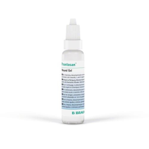
Lo sbrigliamento chirurgico è il metodo preferito per il trattamento delle ulcere del piede diabetico. Esso prevede l'uso di bisturi, forbici o curette per rimuovere il tessuto necrotico fino a quando non si osserva un leggero sanguinamento, che indica la presenza di tessuto vitale. L'efficacia di questa procedura è potenziata dalla previa applicazione di agenti detergenti come Prontosan®.
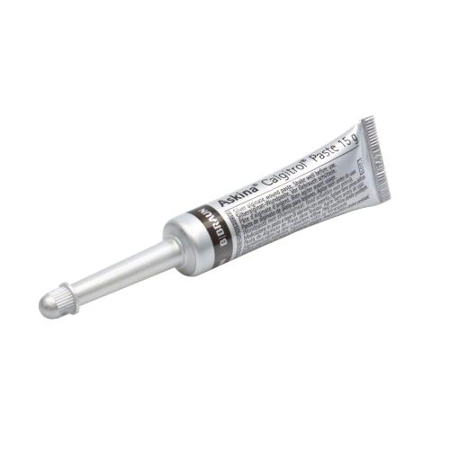
L'uso di medicazioni all'argento previene la riformazione del biofilm e riduce la carica batterica.[7]
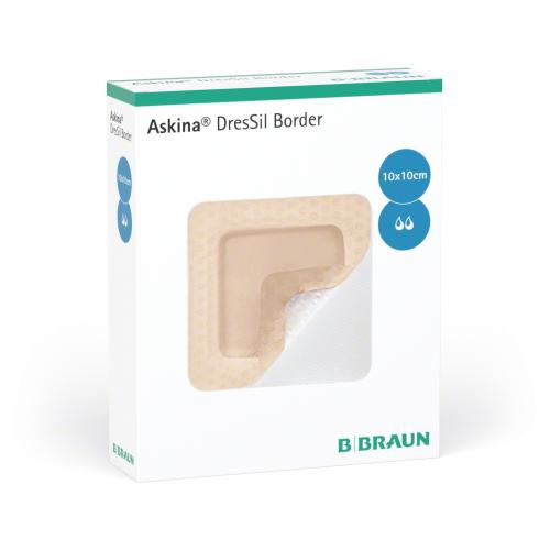
Utilizzare medicazioni adeguate, quali medicazioni in alginato o superassorbenti, per un controllo efficace dell'essudato della ferita. Un'eccessiva umidità può causare la macerazione della cute circostante e ritardare la guarigione.[7]
20 novembre 2025 – 12:00 CEST
Il Prof. Dr. Jose Luis Lázaro Martinez vi guiderà alla scoperta del peso e delle sfide delle ulcere del piede diabetico (DFU) e condividerà approfondimenti e best practice nella gestione delle infezioni attraverso casi clinici reali. I partecipanti otterranno una panoramica completa dalla diagnosi al trattamento, compresi gli approcci terapeutici e le strategie di follow-up dei pazienti.
Registrati ora
[1] Rümenapf G, Morbach S, Rother U, Uhl C, Görtz H, Böckler D, Behrendt CA, Hochlenert D, Engels G, Sigl M; Kommission PAVK und Diabetisches Fußsyndrom der DGG e. V.. Diabetisches Fußsyndrom – Teil 1 : Definition, Pathophysiologie, Diagnostik und Klassifikation [Diabetic foot syndrome-Part 1 : Definition, pathophysiology, diagnostics and classification]. Chirurg. 2021 Jan;92(1):81-94. German. doi: 10.1007/s00104-020-01301-9. PMID: 33170315; PMCID: PMC7819949.
[2] Sommer TC, Lee TH. Charcot foot: the diagnostic dilemma. Am Fam Physician. 2001 Nov 1;64(9):1591-8. Erratum in: Am Fam Physician 2002 Jun 15;65(12):2436-8. PMID: 11730314.
[3] Callaghan BC, Cheng HT, Stables CL, Smith AL, Feldman EL. Diabetic neuropathy: clinical manifestations and current treatments. Lancet Neurol. 2012 Jun;11(6):521-34. doi: 10.1016/S1474-4422(12)70065-0. Epub 2012 May 16. PMID: 22608666; PMCID: PMC4254767.
[4] Takeo M, Lee W, Ito M. Wound healing and skin regeneration. Cold Spring Harb Perspect Med. 2015 Jan 5;5(1):a023267. doi: 10.1101/cshperspect.a023267. PMID: 25561722; PMCID: PMC4292081.
[5] Lauterbach S, Kostev K, Kohlmann T. Prevalence of diabetic foot syndrome and its risk factors in the UK. J Wound Care. 2010 Aug;19(8):333-7. doi: 10.12968/jowc.2010.19.8.77711. PMID: 20852505
[6] Sämann A, Tajiyeva O, Müller N, Tschauner T, Hoyer H, Wolf G, Müller UA. Prevalence of the diabetic foot syndrome at the primary care level in Germany: a cross-sectional study. Diabet Med. 2008 May;25(5):557-63. doi: 10.1111/j.1464-5491.2008.02435.x. Epub 2008 Mar 14. PMID: 18346154.
[7] Strain WD, Paldánius PM. Diabetes, cardiovascular disease and the microcirculation. Cardiovasc Diabetol. 2018 Apr 18;17(1):57. doi: 10.1186/s12933-018-0703-2. Erratum in: Cardiovasc Diabetol. 2021 Jun 11;20(1):120. PMID: 29669543; PMCID: PMC5905152.
[8]. see current IFU Linovera®
[9] see current IFU Linovera® Emulsion
[10] see current IFU Askina® Heel
[11] Olm M, Kühnl A, Knipfer E, Salvermoser M, Eckstein HH, Zimmermann A. Operative Versorgung von Diabetikern mit vaskulären Komplikationen : Sekundärdatenanalyse der DRG-Statistik von 2005 bis 2014 in Deutschland [Operative treatment of diabetics with vascular complications : Secondary data analysis of diagnosis-related groups statistics from 2005 to 2014 in Germany]. Chirurg. 2018 Jul;89(7):545-551. German. doi: 10.1007/s00104-018-0628-z. PMID: 29589075
[12] Narres M, Kvitkina T, Claessen H, Droste S, Schuster B, Morbach S, Rümenapf G, Van Acker K, Icks A. Incidence of lower extremity amputations in the diabetic compared with the non-diabetic population: A systematic review. PLoS One. 2017 Aug 28;12(8):e0182081. doi: 10.1371/journal.pone.0182081. PMID: 28846690; PMCID: PMC5573217.
[13] Jeon BJ, Choi HJ, Kang JS, Tak MS, Park ES. Comparison of five systems of classification of diabetic foot ulcers and predictive factors for amputation. Int Wound J. 2017 Jun;14(3):537-545. doi: 10.1111/iwj.12642. Epub 2016 Oct 10. PMID: 27723246; PMCID: PMC7949506.
[14] Santema TB, Lenselink EA, Balm R, Ubbink DT. Comparing the Meggitt-Wagner and the University of Texas wound classification systems for diabetic foot ulcers: inter-observer analyses. Int Wound J. 2016 Dec;13(6):1137-1141. doi: 10.1111/iwj.12429. Epub 2015 Feb 26. PMID: 25720543; PMCID: PMC7949589.
[15] Wagner FW. The dysvascular foot: a system for diagnosis and treatment. Foot Ankle 1981;2
[16] see current IFU Prontosan® Wound Irrigation Solution
[17] see current IFU Prontosan® Wound Gel X
[18] see current IFU Prontosan® Debridement Pad
[19] see current IFU Askina® Calgitrol Paste
[20] see current IFU Askina® DresSil Border
[21] see current IFU Askina® Calgitrol AG
[22] see current IFU Askina® Sorb
[23] see current IFU Askina® DresSil
[24] see current IFU Askina® Foam
[25] see current IFU Askina® Carbosorb