Nessun risultato di contenuto corrisponde alla tua parola chiave.
Contenuto
Sei uscito con successo.
Non sei ancora registrato?
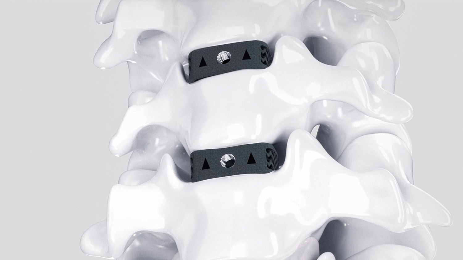
Nuovi spazi per la fusione
Ispirate all'anatomia umana, potenziate dalla scienza, le nostre gabbie fondono i progressi tecnologici con la clinica. Il risultato è un grande balzo in avanti nella stabilizzazione posteriore.
Cliccando "Confirm" dichiari di essere un operatore sanitario.
Se sei un paziente o un giornalista visita le pagine a te dedicate.
Conferma Sì, sono un operatore sanitario. Cancella No, non sono un operatore sanitario.Structan®
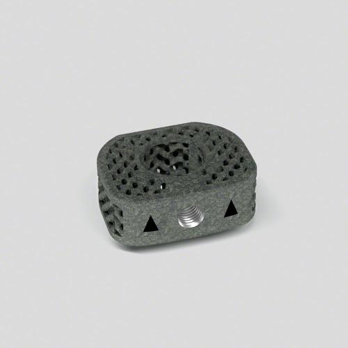
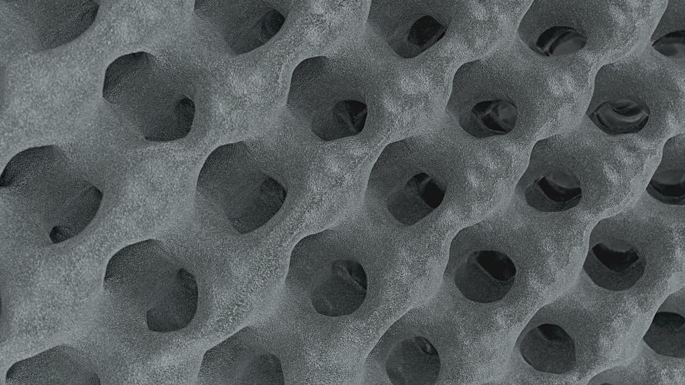
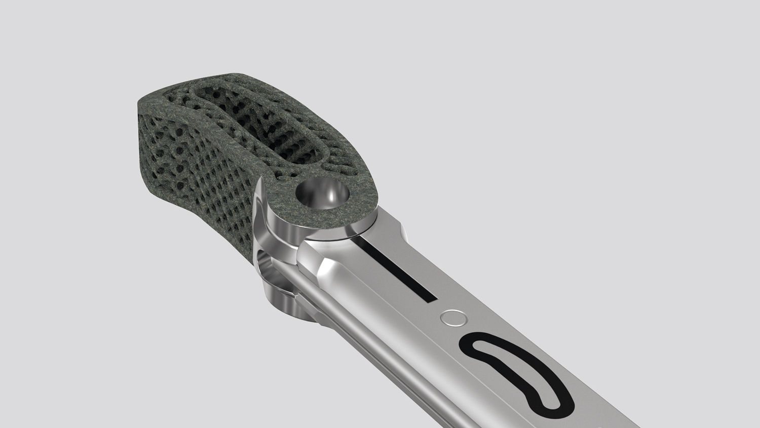
Pensi si tratti di un normale reticolo della gabbia? In questo articolo troverai la scienza alla base di Structan®. Decenni di esperienza, uniti alla tecnologia moderna, hanno portato alla creazione di Structan®, una struttura progettata per dare risultati clinici e prestazioni biomeccaniche avanzate.
La superficie è ingrandita di
0
volte e offre maggiori opportunità di crescita ossea.
Robusto ed elastico allo stesso tempo - Structan® è
0%
più vicino al modulo elastico dell'osso corticale. (1-4) *
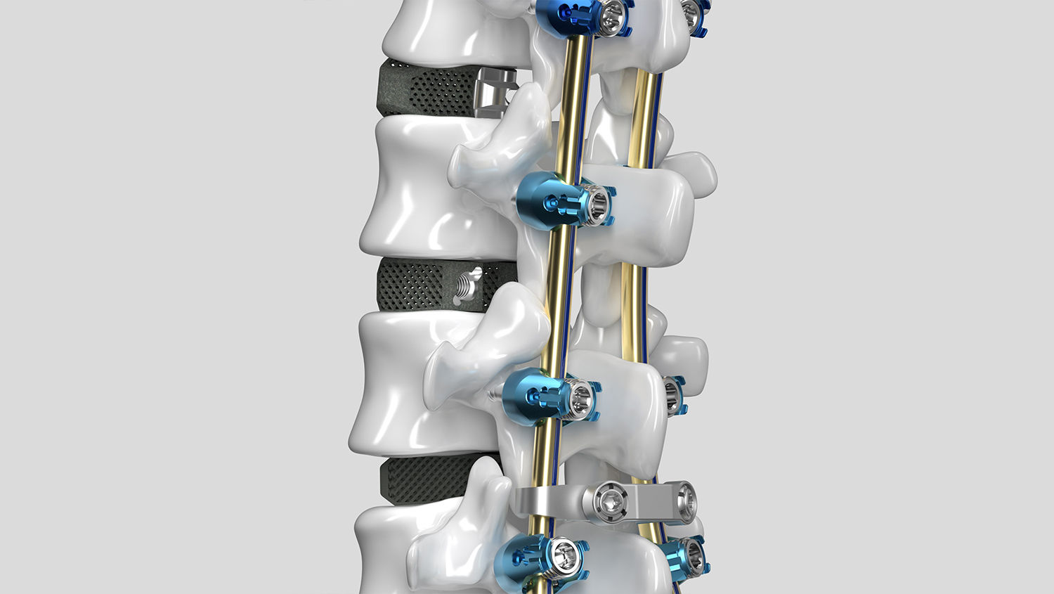
Coprire la stabilizzazione posteriore con
0
piattaforma spinale modulare che si adatta con precisione alle vostre esigenze.
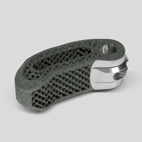
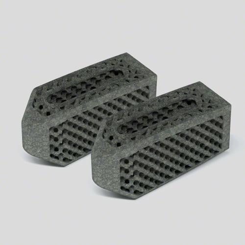
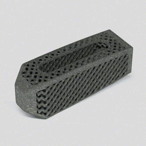
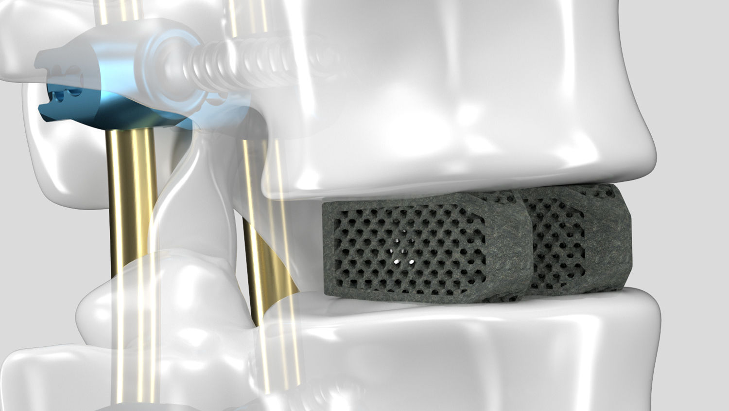
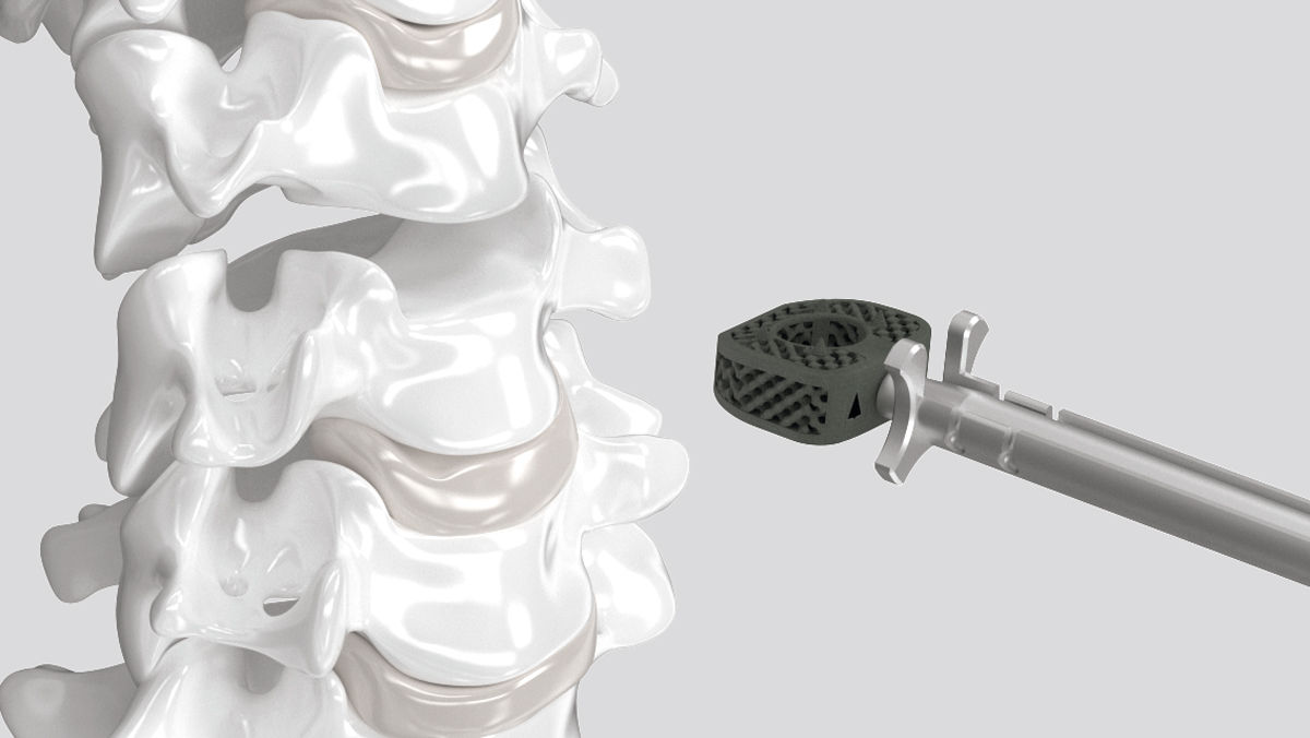
Dispositivi per fusione intersomatica AESCULAP® 3D
Come per la creazione di tutte le nostre soluzioni per la colonna vertebrale, il design dei dispositivi di fusione intersomatica AESCULAP® 3D si basa sui nostri valori fondamentali: creare prestazioni biomeccaniche avanzate, flessibilità intraoperatoria e migliori risultati clinici.
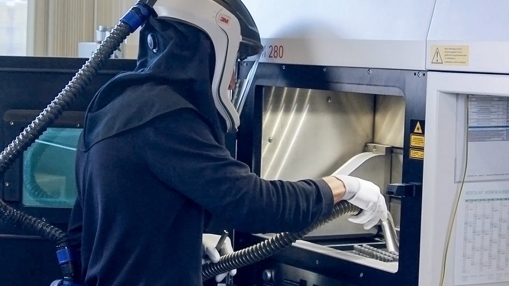
Produzione additiva
Biomeccanica Structan®
Range di misura
Animazioni chirurgiche
Osserva le prestazioni dei nostri dispositivi di fusione intersomatica AESCULAP® 3D.
Il nostro dispositivo di fusione intersomatica TLIF, con il suo introduttore articolato, consente vere procedure mininvasive di fusione.
Grazie alla sua tecnica chirurgica semplificata, i nostri dispositivi di fusione intersomatica PLIF ed Ennovate® sono ideali per l'approccio a cielo aperto.
Unendo l'essenza di due mondi: il nostro dispositivo di fusione intersomatica OLIF può essere impiantato con un approccio mininvasivo e open.
Scopri la piattaforma spinale AESCULAP®
*Rispetto ai dispositivi di fusione intersomatica in lega di titanio massiccio.
[1] Kuhn JL, Goldstein SA, Choi K, London M, Feldkamp LA, Matthews LS. Comparison of the trabecular and cortical tissue moduli from human iliac crests. J Orthop Res. 1989;7(6):876 84.
[2] Ratner BD, Hoffmann AS, Schoen FJ, Lemons JE. An Introduction to Materials in Medicine. Academic Press. 1996.
[3] Chen Y, Wang X, Lu X, Yang L, Yang H, Yuan W, et al. Comparison of titanium and polyetheretherketone (PEEK) cages in the surgical treatment of multilevel cervical spondylotic myelopathy: a prospective, randomized, control study with over 7 year follow up. Eur Spine J. 2013;22(7):1539 46.
[4] Brizuela A, et al. Influence of the elastic modulus on the Osseointegration of Dental Implants. Materials. 2019;12(6):980.
[5] Bostrom M, Boskey A, Kaufman J, Einhorn T. Form and function of bone. In: Orthopaedic Basic Science Biology and Mechanics of the Musculoskeletal System. 2nd ed. Rosemont, IL: AAOS; 2000: 320-369.
[6] Olivares-Navarrete R, Gittens RA, Schneider JM, et al. Rough titanium alloys regulate osteoblast production of angiogenic factors. Spine J 2012; 12:265-272.
[7] Lincks, J. et al. Response of MG63 osteoblast-like cells to titanium and titanium alloy is dependent on surface roughness and composition. Biomaterials 19, 1998. Pages 2219-32.
[8] Elias CN, et al. Mechanical and clinical properties of titanium and titanium based alloys ( Ti G2, Ti G4 cold worked nanostructured and Ti G5) for biomedical applications. Journal of Materials Research and Technology. 2019;8(1):1060 9.
[9] Cheng A, Cohen D, Boyan B et al. Laser-Sintered Constructs with Bio-inspired Porosity and Surface Micro/ Nano-Roughness Enhance Mesenchymal Stem Cell Differentiation and Matrix Mineralization In Vitro. Calcif Tissue Int 2016; 99:625–637.
[10] Wu S-H, Li Y, Zang Y-Q, et al. Porous Titanium-6 Aluminum-4 Vanadium Cage Has Better Osseointegration and Less Micromotion Than a Poly-Ether-Ether-Ketone Cage in Sheep Vertebral Fusion. Art Organs 2013; 37:191-201
[11] Taniguchi N, Fujibayashi S, Takemoto M, Sasaki K, Otsuki B, Nakamura T, Matsushita T, Kokubo T, Matsuda S. Effect of pore size on bone ingrowth into porous titanium implants fabricated by additive manufacturing: An in vivo experiment. Materials Science and Engineering 2016; C59: 690–701.
[12] Changhui Song, Lisha Liu, Zhengtai Deng, Haoyang Lei, Fuzhen Yuan, Yongqiang Yang, Yueyue Li, Jiakuo Yu. Research progress on the design and performance of porous titanium alloy bone implants. Journal of Materials Research and Technology, Volume 23, 2023. Pages 2626-2641, ISSN 2238-7854.
[13] Fukuda A, Takemoto M, Saito T, et al. Osteoinduction of porous Ti implants with a channel structure fabricated by Selective Laser Melting. Acta Biomat 2011; 7:2327-2336.
[14] Ran Q, Yang W, Hu Y, Shen X, Yu Y, Xiang Y, Cai K. Osteogenesis of 3D printed porous Ti6Al4V implants with different pore sizes. J Mech Behav Biomed Mater. 2018 Aug;84:1-11. doi: 10.1016/j.jmbbm.2018.04.010. Epub 2018 Apr 18. PMID: 29709846.
[15] Van Bael S, Chai YC, Truscello S, Moesen M, Kerckhofs G, Van Oosterwyck H, Kruth JP, Schrooten J. The effect of pore geometry on the in vitro biological behavior of human periosteum-derived cells seeded on selective laser-melted Ti6Al4V bone scaffolds. Acta Biomater. 2012 Jul;8(7):2824-34. doi: 10.1016/j.actbio.2012.04.001. Epub 2012 Apr 7. PMID: 22487930.
[16] Kia, C.; Antonacci, C.L.; Wellington, I.; Makanji, H.S.; Esmende, S.M. Spinal Implant Osseointegration and the Role of 3D Printing: An Analysis and Review of the Literature. Bioengineering 2022, 9, 108. https://doi.org/10.3390/bioengineering9030108.
[17] Usability-Test, Usability Validation of AESCULAP® CeSPACE® 3D Cages, Tübingen, 2019.The usability of the AESCULAP® 3D Cage System CeSPACE® 3D was tested in April 2019, in a cadaver workshop with six independent test persons as intended users (surgeons specialized in spinal surgery or comparable fields). Parameters such as implant visibility under x-ray control, mechanical stability of the implant/instrument interface andimplant surface evaluation in terms of tissue injury risk were tested among others. Acceptance criteria were fulfilled for all the above-mentioned parameters. All test users confirmed the absence of critical features that must be improved prior to clinical use. During the test, the x-ray visibility of the cages was particularly positively assessed.
[18] Rehnitz, Christoph, PD Dr. med. Radiological image evaluation of AESCULAP® interbody fusion devices, Heidelberg, 2019. CT and X-ray visualization of different AESCULAP® interbody fusion cages (full titanium, porous Ti6Al4V and PLASMAPOREXP® cages) was tested in a cadaver setup. A radiologist evaluated the implant visibility and the presence of artefacts that may limit the visualization of adjacent structures. Visualization and assessment of implant position was achieved in X-ray and CT for all tested cages. Minor artefacts were visible in CT reconstructions in the surrounding of porous Ti6Al4V and full titanium implants. Porous Ti6Al4V implants showed slightly fewer artefacts in CT in comparison to full titanium implants. The minor artefacts observed did not limit the assessment of the surrounding anatomical structures.